Birth Demonstration Model
Birth Demonstration Model
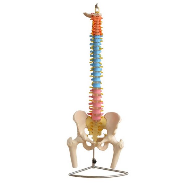
This model consists of 2 innominate bones, sacrum, coccyx, with fetal skull which is firmly connected with the model by means of a flexible metal tube which can be fixed in any position.
Specifications
Size: 58x45x50cm
Material: PVC
Pieces: 8 pieces
Weight: 15kgs
Birth Demonstration Model
Pregnancy and its changes is a normal physiological process that happens in all mammalian in response to the development of the fetus. These changes happen in response to many factors; hormonal changes, increase in the total blood volume, weight gain, and increase in fetus size. All these factors have a physiological impact on all systems of the pregnant woman; musculoskeletal, endocrine, reproductive system, cardiovascular, respiratory, gastrointestinal system, and renal changes.
Anatomy Background
- The pelvis is the region found between the trunk and lower limbs.
- In females, the pelvis is wider and lower than that of their male counterpart, making it more suited to accommodate a fetus during both pregnancy and delivery.
- It protects and supports the pelvic contents, provides muscle attachment and facilitates the transfer of weight from trunk to legs in standing, and to the ischial tuberosities in sitting.
- The cross-sectional anatomy of the female pelvis shows five bones: two hip bones, sacrum, coccyx, and two femurs. The joints are supported by some of the strongest ligaments in the body which become laxer during pregnancy leading to increased joint mobility and less efficient load transfer through the pelvis.
- The pelvic outlet at the base of the pelvis is narrower in its transverse diameter when compared with the pelvic inlet; it comprises the pubic arch, ischial spines, sacrotuberous ligaments, and coccyx.
Musculoskeletal Changes
- The overall equilibrium of the spine and pelvis alters as the pregnancy progresses
- There is still confusion as to the exact nature of any associated postural adaptation. With weight gain, increased blood volume, and ventral growth of the fetus,
- The centre of gravity no longer falls over the feet, increase in anteroposterior and medial-lateral sway, and women may need to lean backwards to gain equilibrium resulting in disorganisation of spinal curves.
- Reported postures include a reduction in lumbar lordosis an increase in both lumbar lordosis and thoracic kyphosis or a flattening of the thoracolumbar spinal curve.
- There will be compensatory changes to posture in the thoracic and cervical spines, and this combined with the extra weight of the breasts may result in posterior displacement of the shoulders and thoracic spine, increase anterior pelvic tilting, and increase of the cervical lordosis.


 Botzees
 Keyestudio
 Fischertechnik
