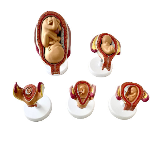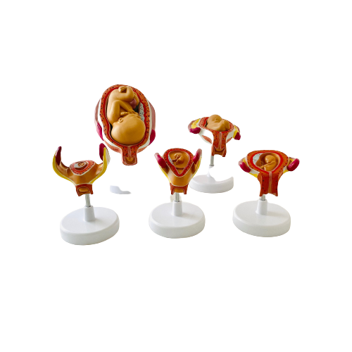
 Botzees

Lung Model
Lung Model
A life-size model separating into 4 parts. The lungs have two removable lobes to show the internal of lung. Life-size, cutaway lung model shows bronchus, arteries, veins, lymph nodes, bronchial passage and trachea bifurcation.
Size: 57x46x62cm
Material: PVC
Life Size Model
Lung Model
The lungs are the primary organs of the respiratory system in humans and many other animals including a few fish and some snails. In mammals and most other vertebrates, two lungs are located near the backbone on either side of the heart. Their function in the respiratory system is to extract oxygen from the atmosphere and transfer it into the bloodstream, and to release carbon dioxide from the bloodstream into the atmosphere, in a process of gas exchange. Respiration is driven by different muscular systems in different species. Mammals, reptiles and birds use their different muscles to support and foster breathing. In early tetrapods, air was driven into the lungs by the pharyngeal muscles via buccal pumping, a mechanism still seen in amphibians. In humans, the main muscle of respiration that drives breathing is the diaphragm. The lungs also provide airflow that makes vocal sounds including human speech possible.
Humans have two lungs, a right lung, and a left lung. They are situated within the thoracic cavity of the chest. The right lung is bigger than the left, which shares space in the chest with the heart. The lungs together weigh approximately 1.3 kilograms (2.9 lb), and the right is heavier.
Lung Model
Function
The major function of the lungs is gas exchange between the lungs and the blood. The alveolar and pulmonary capillary gases equilibrate across the thin blood–air barrier. This thin membrane (about 0.5 –2 μm thick) is folded into about 300 million alveoli, providing an extremely large surface area (estimates varying between 70 and 145 m2) for gas exchange to occur.

The effect of the respiratory muscles in expanding the rib cage.
The lungs are not capable of expanding to breathe on their own, and will only do so when there is an increase in the volume of the thoracic cavity. This is achieved by the muscles of respiration, through the contraction of the diaphragm, and the intercostal muscles which pull the rib cage upwards as shown in the diagram. During breathing out the muscles relax, returning the lungs to their resting position. At this point the lungs contain the functional residual capacity (FRC) of air, which, in the adult human, has a volume of about 2.5–3.0 litres.